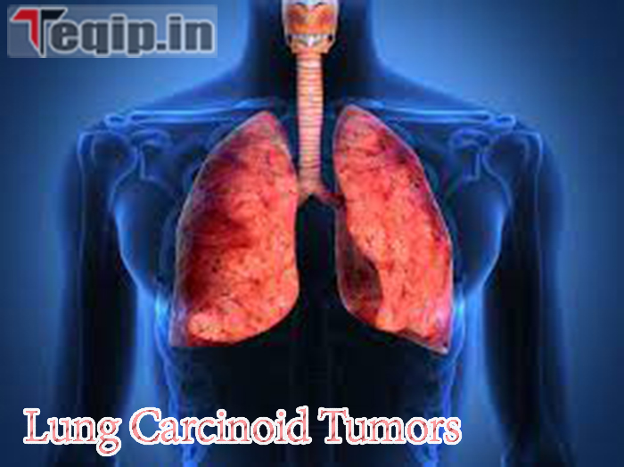
Lung Carcinoid Tumors:- Carcinoid tumors are a very rare form of lung cancer. Also known as pulmonary neuroendocrine tumor (NET), it accounts for only one to two percent of all lung cancers. It grows slowly. It is so named because it starts growing in certain specialized cells called neuroendocrine cells. These cells are found throughout the body. In the early stages, it usually causes no symptoms and is detected during testing for another disease or surgery.
When symptoms do appear, they can include tiredness, nausea, or pain, and signs such as fever, a rash, or a rapid heart rate. Together, the signs and disorder can help describe a medical problem. It too define the signs and disorder of carcinoid syndrome & carcinoid crisis, that are conditions that canister cause pulmonary NETs. If you knowledge any of these signs and symptoms, you allow consult your doctor.
Lung Carcinoid Tumors
Neuroendocrine cells are found throughout the body. The Lung Carcinoid Tumors consists exclusively of these neuroendocrine cells. These cells are so named because they resemble endocrine cells and secrete hormone-like substances.
On the other hand, like nerve cells, they are also able to secrete neurotransmitters. Under unusual circumstances, the growth rate of these nerve cells increases and tumors form. Such tumors can form anywhere in the body. In fact, lung carcinoids account for only about 2% of all-body cancers in the same category.
Lung Carcinoid Tumors Details
| Article Name | Lung Carcinoid Tumors |
| Category | Health |
| Official Website | Click Here |
Check also:- Covid XBB 1.5 Variant
Types of Lung Carcinoid
Typical Lung Carcinoids
90% of Lung Carcinoid Tumors are of the typical type and are found in the central airways of the lungs. Their growth rate is slow compared to other lung tumors and rarely extends beyond the lungs. It resembles a malignant lung cancer with signs and symptoms such as a severe cough and hemoptysis. Another way to categorize pulmonary NETs is by location. Those located on the outer edge are called peripheral carcinoids. And those located in the middle of the airways are called central carcinoids. About 80-90% of patients live another 5 years with this carcinoid.
Atypical Lung Carcinoids
They are rarer than typical carcinoids and can metastasize beyond the lungs. They also tend to grow faster than typical carcinoids. These tumors show an increased activity of cell division and cell death (necrosis). About 50-70% of patients live with this cancer for another 5 years.
Causes of Lung Carcinoids
The exact cause of Lung Carcinoid Tumors forming before any other lung cancer is not very certain, however certain factors that increase risk may include:
Gender
Women are more prone to lung cancer than men.
Skin Color
It is more common in people of white race than in any other people of any race.
Genetic
People with a family history or who inherited the gene for MEN-1 (multiple endocrine neoplasia type 1) have a higher risk of developing lung carcinoids than others. The presence of this syndrome acts as a precursor to other carcinoids such as pancreatic carcinoids, pituitary, etc.
Family History
If carcinoids run in your family, you are at a higher risk of being caught.
Read also:- Pregnancy Week 15 Baby
Lung Carcinoids Symptoms
Usually, there are no specific symptoms of lung carcinoid and it is usually identified by imaging, scanning, etc. during diagnosis of other patient ailments or during chest examination. Symptoms generally include:
Cough And Wheezing
It may contain sputum and sometimes sputum along with blood. In severe cases.
Infections
When the tumor grows so large that it blocks the airway, post-obstructive pneumonia occurs.
Carcinoid Syndrome
It is very rare, but when it does, the neuroendocrine cells of the tumor start overproducing certain hormones.
Facial Flushing
It usually resembles blushing and is the redness of the face. However, in doing so, the person feels the warmth on their face, a burning hot sensation that is due to the overproduction of serotonin. In this state, serotonin is overproduced and released into the systemic circulation.
Hirsutism
an excessive growth of facial and body hair.
Diagnosis For Lung Carcinoids
If you experience any of the above symptoms, you should consult a pulmonologist or a general practitioner. They will ask you about your family history and will first physically examine your lungs. A series of tests are then performed to examine the issue in detail. This could include:
Blood Test
A blood test and urine test are done to look for abnormal or elevated hormones or substances. It looks for the presence of serotonin and chromogranin A. The presence of this compound indicates a carcinoid tumor.
Urine Test
It is performed to look for the serotonin metabolite 5-HIAA. Both tests provide reproducible results for people with carcinoid syndrome and pulmonary carcinoid. But for people without symptoms, these tests are quite sufficient.
X-ray
An X-ray will detect the presence of a tumor in the lungs unless the tumor is short or covered by another organ.
CT-Scan
It is a 3D imaging technique. If an X-ray does not provide clarity, it can be done to find even the smallest tumor and the exact location of the tumor. It can even indicate the spread status of the tumor.
Biopsy
This is a process in which a tiny part of the tumor is removed and examined under a microscope. There are two kinds:
Surgical
It is like an operation in which a cavity is created in the chest under the influence of anesthesia and a tumor sample is then taken. This requires adequate recovery time and hospitalization.
Non-surgical
This happens in normal hospitals or clinics. A sample is taken under the influence of a normal sedative without a surgical incision. The best example of this is bronchoscopy, where a thin wire is inserted through the windpipe into the chest and a biopsy is taken under direct vision.
Check this:- Zika Virus Symptoms
Lung Carcinoids Stages
The stages of Lung Carcinoid Tumors are identified based on the size of the tumor (T), its spread to surrounding lymph nodes (N), and metastasis (M). It is divided into four stages. Stage zero is the smallest tumor and stage four is the largest sized tumor. The further classification of lung carcinoid tumors according to AJCC (American Joint Committee on Cancer) is shown in the table below.
| S.NO. | AJCC Staging | Stage Group | Description |
| 1 | occult cancer | TX | the cancer is found somewhere in the body but cannot located. |
| NO | cancer has not spread to the surrounding lymph node | ||
| MO | cancer has not spread to any more body part | ||
| 2 | 0 | Tis | the cancer cells are found superficially on the air passage and have not invaded deeper |
| NO | cancer has not spread to the surrounding lymph node | ||
| MO | cancer has not spread to any more body part | ||
| 3 | IA1 | T1a | The tumor has not reached the main branches of the bronchi and the membrane of the lungs and is no larger than 1 cm in size. |
| NO | cancer has not spread to the surrounding lymph node | ||
| MO | cancer has not spread to any other body part | ||
| 4 | IA2 | T1b | The tumor has not reached the main branches of the bronchi and the membrane of the lungs and is approx. 1-2 cm in size. |
| NO | cancer has not spread to the surrounding lymph node | ||
| MO | cancer has not spread to any other body part | ||
| 5 | IA3 | T1c | The tumor has not reached the main branches of the bronchi and the membrane of the lungs and is approx. 2-3 cm in size. |
| NO | cancer has not spread to the surrounding lymph node | ||
| MO | cancer has not spread to any other body part | ||
| 6 | IB | T2a | 1)The crab is 3-4 cm in size. 2) The tumor has grown into the main branches of the bronchi and visceral pleura, partially obstructing the air passage, but has not reached the carina |
| NO | cancer has not spread to the surrounding lymph node | ||
| MO | cancer has not spread to any other body part | ||
| 7 | IIA | T2b | 1) The crab is 4-5 cm in size. 2) The tumor has grown into the main branches of the bronchi and visceral pleura and partially obstructs the airways and has also reached the carina. |
| NO | cancer has not spread to the surrounding lymph node | ||
| MO | cancer has not spread to any other body part | ||
| 8 | IIB | T | T1a/T1b/T1c or T1a/T2b or T3 |
| T3 | The tumor is 5-7 cm in size and has now spread to the parietal pleura and the parietal pericardium. There’s more than one tumor in the lungs. | ||
| NO | NO or N1 | ||
| N1 | cancer has spread to the hilar lymph node and lymph node and is on the same side | ||
| MO | cancer has not spread to any other body part | ||
| 9 | III A | T | T1a/T1b/T1c or T1a/T2b or T3 or T4 |
| T3 | The tumor is 5-7 cm in size and has now spread to the parietal pleura and the parietal pericardium. There’s more than one tumor in the lungs. | ||
| T4 | The tumor is larger than 7 cm and has spread to the heart, blood vessels, trachea, stomach, esophagus and spine. There are more than two tumors in different parts of the lungs. | ||
| N | N2 or N3 | ||
| N2 | The tumor has spread to the carina and the space between the lungs. Lymph nodes and tumor are on the same side | ||
| N3 | Cancer has spread to the collarbone on either side of the body. | ||
| Mo | cancer has not spread to any other body part | ||
| 10 | III B | T1a/T1b/T1c, N3,MO | |
| T2a/T2b, N3, MO | |||
| T3,N2.MO | |||
| T4,N2,MO | |||
| 11 | III C | T3,N3,MO | |
| T4,N3,MO | |||
| 12 | IV A | T | any T |
| N | any N | ||
| MO | M1a/ M1b | ||
| M1a | cancer has spread in both the lungs | ||
| M1b | The single tumor has spread outside the lungs to the bone, liver, or brain. | ||
| 13 | IV B | T | any T |
| N | any N | ||
| M1c | more than one tumor has spread to the other organs outside the breast |
A patient’s treatment is based on the diagnosis, which includes the type of tumor (typical or atypical/peripheral or central), whether it has spread to the lungs or other organs, and the stage of the tumor gives. The stage of the tumor is determined by the size of the tumor. The AJCC categorizes the tumor into stages from a minimum as Stage 0 to a maximum as Stage IV.
Treatment includes surgery and radiation. However, most lung carcinoids can be removed with surgery alone. However, if the tumor has spread to other organs, a palliative tumor is removed through surgery to relieve symptoms. Radiation therapy is then given to finally remove it from the body.
Read this:- Digital Health ID Card 2024
Surgical Treatment
Lobectomy
it’s the removal of a love from the lungs. The removal of both lobes is called a bilobectomy. This is done for peripheral tumors. For example, sleeve resection involves removing the tumor along the top and bottom of the airway, and then suturing the airway.
Sulobar Resection
These include segmentectomy, in which the segment of the lobe of the lung is removed. This includes wedge resection, in which a small wedge-shaped part is removed from the lung.
Pneumonectomy
In this procedure, Lung Carcinoid Tumors the entire lung is removed while the tumor spreads to the entire lung.
Dissection Of Lymph Node
When removing a tumor, lymph nodes are often removed to observe the spread of the tumor within them. This is also fixed to avoid the spread of tumors to other organs.
Radiation Treatment
it requires more than one session. High-intensity radiation exposure is used to destroy cancer cells. It is the general therapy for destroying cancer cells. Mall pellets and rods are sent via a catheter to a tumor and then exposed to a beam of radiation to destroy the tumor. This procedure is called brachytherapy.
Chemotherapy
This is the intravenous delivery of drugs into the veins to destroy the cancer cells. Certain medications used in this technique are given as pills, injections, shots, creams, etc. This therapy can be administered in a doctor’s office or in a hospital. However, there are several side effects associated with this treatment, which in many cases are manageable.
Lung Carcinoid Tumors FAQ’S
How often do carcinoid lung tumors survive?
The 10-year endurance in patients with an abnormal carcinoid was 49%, versus the 84% 10-year endurance rate saw in patients with a commonplace carcinoid.
Are carcinoid growths cellular breakdown in the lungs?
Lung cancer of the carcinoid type is uncommon. Carcinoid tumors make up only one to two percent of lung cancers. They normally develop gradually. They are a type of neuroendocrine tumor, which means that they start in neuroendocrine cells, which are special cells that are found in the body and in the lungs.
Where are lung carcinoid tumors located?
In the walls of the large airways (bronchi), which are close to the center of the lungs, central carcinoids develop. The majority of lung carcinoid tumors are typical carcinoids or central carcinoids.
Related Post:-
